
Dog Limping: Causes, First Aid, and Treatment 2025
Dog limping ✓ All causes from harmless to serious ✓ First aid tips ✓ When to see the vet ✓ Assess lameness correctly ✓ Veterinarian reviewed ✓ [Learn more]
⚖️ Legal Notices
Important Information: This article is for general information and education purposes only. It does not replace individual veterinary advice, diagnosis, or treatment.
Veterinary Consultation: If your dog has health problems, always consult a veterinarian. Only a veterinarian can make a correct diagnosis and recommend appropriate treatment.
Dietary Supplements: The dietary supplements mentioned in this article are not a substitute for veterinary treatment and cannot replace such treatment. Dietary supplements are not a substitute for a balanced diet.
Professional Basis and Update
Scientific Basis: This guide is based on veterinary orthopaedic guidelines and specialist literature from:
- German Society for Small Animal Medicine (DGK-DVG) - Professional standards in Germany
- University of Veterinary Medicine Hannover - Small Animal Clinic, Orthopaedics Department
Source Transparency:
- Publication period: 2010-2022
- All studies verifiable via PubMed (PMID numbers linked in the text)
Update: January 2025
What Does Limping in Dogs Mean: Definition and Assessment
When a dog is limping, it shows an abnormal gait due to pain, injury, or disorders of the musculoskeletal system. Limping can range from slight lameness to complete weight-bearing avoidance and, depending on severity, requires different measures - from observation to an immediate visit to the veterinarian.
⚡ QUICK ANSWER: Dog Limping - What to do NOW?
In the first 5 minutes:
- Leash immediately - Stop further movement
- Check the paws - Look for foreign bodies between the toes/pads
- Assess severity - Is the dog still putting weight on the leg?
When to see the vet IMMEDIATELY (within 1 hour):
- The leg is not being used at all (complete weight-bearing avoidance)
- Visible injury with bleeding
- The dog whines when lightly touched
- Swelling or deformity is visible
When to see the vet within 24 hours:
- Limping remains unchanged after 4-6 hours of rest
- Mild to moderate lameness without improvement
- No obvious foreign body found
When 48 hours of observation is OK:
- Only slight limping (the leg is still being used)
- No visible injury
- Improvement with rest is noticeable
- Dog is eating and drinking normally
Read on for detailed diagnostics and treatment options ↓ TL;DR - The most important points: • Limping is always a sign of pain • Distinguish: front leg vs. hind leg, acute vs. chronic • See the vet immediately: with complete weight-bearing avoidance, visible injuries • First aid: rest, check paws, restrict movement • Common causes: injuries, osteoarthritis, foreign bodies, overuse
Table of Contents
- Types of limping
- Common causes by location 2.5. Why is my dog limping? - Most common questions
- Assessing severity correctly
- First aid measures
- When to see the vet immediately
- Examination and diagnostics
- Treatment options
- Home care and support
- Prevention of lameness
- Long-term movement health
Acute vs. chronic lameness
Veterinary specialist literature defines standardized assessment criteria for different forms of lameness in dogs. Acute lameness (sudden onset):
- Duration: Less than 24-48 hours
- Causes: Trauma, injury, foreign body
- Characteristics: Marked pain, often one-sided
- Prognosis: Usually good with appropriate treatment Chronic lameness (long-lasting):
- Duration: More than 2-3 weeks
- Causes: Osteoarthritis, dysplasia, tumors
- Characteristics: Gradual worsening
- Course: Often progressive, requires management Intermittent lameness:
- Pattern: Fluctuating, not constant Typical for: Early osteoarthritis, overuse
- Triggers: Cold, exertion, prolonged lying down
- Assessment: Can be misleading, often underestimated
Front Leg vs. Hind Leg Lameness
Gait Analysis: Recognizing front leg lameness:
- Head movement: Head rises when the painful leg is placed on the ground
- Weight shift: More load on the healthy front leg
- Stride length: Shortened steps on the affected side
- Turning: Difficulty changing direction
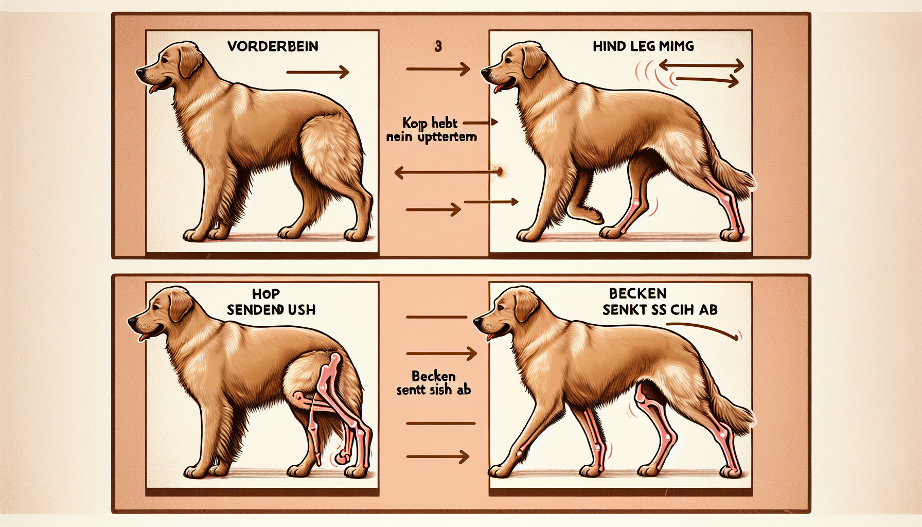
Figure: Gait analysis to distinguish front leg and hind leg lameness. Left: In front leg lameness, the head rises when the affected leg bears weight. Right: In hind leg lameness, the pelvis on the affected side drops.
Recognizing hind leg lameness:
- Pelvic drop: The hip drops on the healthy side
- "Bunny hopping": Both hind legs move together while running
- Difficulty standing up: Trouble getting up
- Stairs: Problems going upstairs
Lameness grades according to AAEP
Modified American Association of Equine Practitioners Scale for Dogs:
| Grade | Description | Weight bearing | Activity |
|---|
[IMAGE NEEDED: Lameness Severity Infographic - Visual guide to assessing lameness from Grade 0 to Grade 5 with action recommendations for each grade]
Paws and Toes
Most common paw problems: Clinical data analyze lameness cases and identify paw problems as a frequent cause.
Objective gait analysis studies (PMID: 33320889) with 41 healthy dogs and 21 dogs with osteoarthritis show characteristic pressure patterns: dogs with osteoarthritis reduce weight bearing by an average of 15-25%, which can be measured precisely using pressure-sensitive walkways. Foreign bodies in the paw: Typical objects: Thorns, glass shards, small stones, awns
- Location: Between the toes, embedded in the pads
- Symptoms: Sudden lameness, licking, swelling Pad injuries:
- Tears and cuts: Sharp objects, rough surfaces
- Burns: Hot asphalt, road salt in winter
- Abrasions: Excessive strain on hard ground
- Symptoms: Bleeding, swelling, pain on touch Nail problems:
- Ingrown nails: Especially dewclaws
- Broken nails: Trauma, nails that are too long
- Nail bed inflammation: Bacterial infection
- Tumors: Squamous cell carcinoma, melanoma (rare)
Hock and Lower Leg
Hock pathologies: Osteochondritis dissecans (OCD):
- Affected breeds: Rottweiler, Labrador Retriever, Great Dane
- Age: 6-18 months
- Pathology: Cartilage-bone lesion
- Symptoms: Lameness after exercise, joint swelling Achilles tendon injury:
- Mechanism: Sudden overextension, chronic overuse
- Symptoms: Hyperflexion of the hock, "plantigrade" stance
- Prognosis: Variable, often requires surgical treatment
- Rehabilitation: 6-12 weeks of immobilization
Knee and Thigh
Cruciate ligament rupture (most common knee injury): Scientific research (PMID: 21243176) identifies cruciate ligament disease as the most common cause of hind leg lameness in dogs. The disease leads to gradual degeneration of the cruciate ligament matrix with eventual ligament rupture. Epidemiology:
- Risk factors: Overweight, age >5 years, certain breeds
- Sex: Neutered animals are more commonly affected Development of symptoms:
- Acute rupture: Sudden lameness, swelling
- Partial rupture: Intermittent lameness
- Chronic: Progressive osteoarthritis
- Meniscal damage: Additional "clicking" sounds Patellar luxation (kneecap luxation):
- Grades 1-4: From occasional to permanently luxated
- Breeds: Small breeds (Chihuahua, Yorkshire Terrier)
- Symptoms: "Skipping" gait, hopping
- Treatment: Conservative (Grade 1-2) or surgical (Grade 3-4)
Hip and Pelvis
Hip dysplasia:
- Genetic: Polygenic inheritance
- Environmental factors: Nutrition, exercise during youth
- Symptoms: Difficulty standing up, "bunny-hopping" gait
- Diagnosis: X-ray, hip dysplasia examination Legg-Calvé-Perthes disease:
- Affected: Small breeds, 6-12 months old
- Pathology: Aseptic necrosis of the femoral head
- Symptoms: Progressive lameness, muscle wasting
- Treatment: Usually surgical removal of the femoral head
💬 Veterinary Consensus on Lameness Treatment
According to veterinary orthopaedic literature and recommendations from the German veterinary profession, the following guidelines apply to lameness in dogs:
Important time windows according to veterinary standards:
- Lameness >24 hours without improvement → veterinary evaluation recommended
- Sudden lameness after trauma → prompt veterinary examination
- Gradual worsening over weeks → osteoarthritis diagnostics advisable
Why is My Dog Limping? Most Common Questions
"My dog is suddenly limping - is that dangerous?" A limping dog is always showing pain or discomfort in the musculoskeletal system. Sudden limping usually indicates an acute injury - for example from a foreign body in the paw, a strain, or trauma. Gradual limping over weeks, on the other hand, is more likely to develop from chronic causes such as osteoarthritis or degenerative joint disease.
"Dog is lame after a walk - normal?" If your dog starts limping after longer walks or intense exercise, several causes are possible: muscle overload, early joint problems, or overexertion in untrained dogs. If the pattern recurs after exercise, you should consult a veterinarian to rule out early joint changes.
"Why is my old dog limping?"
"Dog limping after getting up - what does that mean?" Stiffness and limping after getting up ("start-up lameness") is characteristic of early or existing osteoarthritis. The joints are less mobile after rest periods, which improves with the first movements. This should be checked by a veterinarian, because early intervention can positively influence the course of the disease.
Emergency Indicators
Immediate veterinary emergency: Absolute emergency signs:
- Complete weight-bearing avoidance: The leg is not used at all
- Visible fractures: Open fractures, deformities
- Severe bleeding: Arterial bleeding from wounds
- Shock symptoms: Pale mucous membranes, weak pulse
- Loss of consciousness: After trauma or a fall Urgent cases (within 2-4 hours):
- Severe lameness Grade 4-5: Little or no weight bearing
- Swelling with heat: Signs of acute inflammation
- Fever: Body temperature >39.5°C
- Persistent pain: No improvement despite rest
- Neurological deficits: Signs of paralysis
Pain Assessment in Dogs
Canine Pain Assessment Tools:
- Physiological parameters: Heart rate, respiratory rate
- Behavioral parameters: Activity, attention, vocalization
- Interaction response: Reaction to touch
- Score 0-27: <6 no pain, >13 severe pain Glasgow Pain Scale (Short Form):
- Mobility: Assessment of movement
- Attention: Interest in surroundings
- Vocalization: Spontaneous or when touched
- Score 0-24: <5 mild, 5-10 moderate, >10 severe Behavioral indicators of pain:
- Panting: Without exertion or heat
- Restlessness: Cannot find a comfortable position
- Loss of appetite: Refuses favorite food
- Aggression: Unusual defensive reactions
- Hiding: Retreat to quiet places
Documentation for the Vet
Record important observations: History information:
- Onset: When first noticed? Sudden or gradual?
- Activity: What was the dog doing beforehand?
- Course: Improvement or worsening?
- Triggers: Specific movements or situations? Video documentation:
- Gait: Film slowly walking from the side
- Different surfaces: Grass, asphalt, stairs
- Getting up/lying down: Document movement sequences
- Duration: 30-60 seconds per sequence
📹 Video Guide: Assessing Gait Correctly
[VIDEO NEEDED: 3-minute video "How do I recognize lameness in a dog?"]
This video shows how you can:
- Film your dog's gait
- Distinguish front leg vs. hind leg lameness
- Assess severity
Immediate Steps
First aid protocol for lameness: 1. Rest and safety:
- Put the dog on a leash immediately
- Create a quiet environment
- Stop all further activity
- Minimize stress signals 2. Visible injuries:
- Carefully inspect the paws
- Do NOT remove foreign bodies yourself (except superficially)
- Stop bleeding with a clean cloth
- Do not rinse wounds (it obscures findings) 3. Paw check:
- Look between the toes
- Check pads for cuts/tears
- Check nails for breaks
- Feel for swelling 4. Pain relief:
- ❌ Do NOT give human painkillers!
- Cool with a damp cloth (not ice directly)
- Quiet, padded surface
- Speak calmly and reassuringly
Transport Safety
Gentle transport to the vet: Small dogs (up to 15kg):
- In a stable transport box
- Soft padding for the affected leg
- Secure the box in the car
- Companion for reassurance Large dogs (over 15kg):
- Minimize movement as much as possible
- For hind leg problems: harness instead of collar
- Use a ramp or carrying basket into the car
- Second person to help Transport avoidance:
- Do not force the dog to walk
- No long walks "to test it"
- Avoid stairs and jumps
- In severe pain: consider a home visit by the vet
Cooling and Stabilization
Thermotherapy in the acute phase: Cold application (first 24-48 hours):
- Cold packs: Never directly on the skin
- Towel layer: Protection against frostbite
- Application time: 10-15 minutes every 2 hours
- Effect: Reduction in swelling, pain relief
Absolute Emergency Situations
Immediate veterinary help required: Traumatic injuries:
- Open fractures: Bone breaks through the skin
- Luxations: Visible joint dislocations
- Severe bleeding: Unstoppable bleeding
- Deep wounds: Tendons or muscles visible
- Foreign body penetration: Object stuck in the paw Neurological symptoms:
- Signs of paralysis: Leg cannot be moved
- Loss of reflexes: No response to touch
- Coordination disorders: Ataxia, staggering
- Altered consciousness: Occurred after trauma
Urgent Cases (within 4-6 hours)
Severe lameness without visible injury:
- Complete or almost complete weight-bearing avoidance
- Severe pain on light touch
- Persistent vocalization (whining, whimpering)
- Fever in combination with lameness Progressive worsening:
- Lameness becomes worse despite rest
- Swelling increases rapidly
- Dog refuses any movement
- Systemic symptoms (vomiting, lethargy)
Observation vs. Treatment
When to observe for 24-48 hours: Mild lameness (Grade 1-2) with:
- Normal weight bearing
- No visible injuries
- No fever
- Normal activity between movements
- Improvement with rest Observation protocol:
- Restrict movement strictly
- Assess condition every 4-6 hours
- Document pain behavior
- If worsening, go to the vet immediately
💰 Cost Transparency: Diagnostics at the Vet
Important upfront: Costs can vary depending on region, veterinarian, and scope of examination. The German schedule of veterinary fees (GOT) regulates the fee ranges in Germany.
Diagnostic Costs at a Glance
| Examination | Base rate (GOT) | 2-3x rate (common) | Emergency service (up to 4x) |
|---|---|---|---|
| General examination | €13.47 | €26.94 - €40.41 | up to €53.88 |
| Lameness examination | €31.66 | €63.32 - €94.98 | up to €126.64 |
| X-ray (2 images) | €44.88 | €89.76 - €134.64 | up to €179.52 |
| Ultrasound | €31.89 | €63.78 - €95.67 | up to €127.56 |
| Joint puncture | €46.01 | €92.02 - €138.03 | up to €184.04 |
| CT examination | €380-€600 | €400-€800 | €800-€1,200 |
| MRI examination | €700-€1,000 | €800-€1,500 | €1,200-€2,000 |
| Blood test (basic) | €22.52 | €45.04 - €67.56 | up to €90.08 |
| Arthroscopy (surgery) | €800-€1,500 | €1,200-€2,500 | €2,000-€3,500 |
Notes on cost estimates:
- Base rate: Rare in practice, usually only for simple routine examinations
- 2-3x rate: Standard during normal consultation hours, reasonable time expenditure
- Emergency service/weekend: Up to 4x the base rate possible, legally permitted
- Additional costs: Sedation (€30-€80), anesthesia (€80-€200), contrast agent (€40-€100) not included
Example Cost Breakdown: Acute Lameness
Scenario 1: Simple injury (foreign body in paw)
- General examination: €30
- Foreign body removal: €40
- Bandage: €25
- Pain medication (3 days): €15
- Total: approx. €110
Scenario 2: Suspected cruciate ligament tear
- Extended lameness examination: €90
- X-ray (2 views): €120
- Sedation: €50
- Pain medication + anti-inflammatory: €35
- Total: approx. €295
Scenario 3: Complex diagnostics (hip dysplasia)
- Special examination: €120
- Hip X-ray (special positioning): €180
- CT examination (both hips): €650
- Anesthesia: €150
- Follow-up examination: €30
- Total: approx. €1,130
💡 Cost-Saving Tips
Pet insurance:
- Surgery insurance: Covers major procedures (€20-€40/month)
- Full coverage: Includes diagnostics (€50-€100/month)
- Tip: Take out insurance while young (lower premiums)
Direct route to the specialist:
- Ask for a referral in chronic cases
- Get a second opinion (can save costs)
Regional differences:
- City: Usually higher fee rates (3x rate common)
- Rural areas: Often 2x rate is sufficient
- Veterinary clinics vs. private practices: Clinics are usually more expensive, but offer more comprehensive diagnostics
⏱️ Timeline: From Symptom to Diagnosis
Realistic timing helps manage expectations and choose the right treatment path.
Day 1: Symptom noticed
Immediate measures (first hour):
- ✅ Restrict movement (leash, no play)
- ✅ Check paws and legs for injuries
- ✅ Assess severity (Grade 1-5 scale)
- ✅ Document behavior (photo/video)
Decision:
- Grade 4-5 (severe lameness): GO TO THE VET IMMEDIATELY (within 1-2 hours)
- Grade 2-3 (moderate lameness): Arrange an appointment for the next day
- Grade 1 (mild): 24-48h rest + observation
Day 1-3: Observation phase (for mild lameness)
What you should do:
- Strict leash walking only (5-10 min walks only)
- Check condition every 6 hours
- Keep a diary: better/same/worse?
- No self-medication (no ibuprofen!)
Warning signs -> Go to the vet immediately:
- Worsening despite rest
- Swelling increases
- Fever (>39.5°C rectal)
- Refusal to eat
Day 3-7: Veterinary diagnostics
Initial appointment (30-45 minutes):
- History: When, how, where did it start?
- Gait analysis: walking on hard ground
- Palpation: locate pain points
- Joint mobility: range of motion tests
- Cost: €60-€100 (examination + consultation)
Imaging (if required):
-
X-ray (standard case):
- Appointments usually same/next day
- Duration: 20-30 minutes
- Result: Can be discussed immediately
- Cost: €120-€180
-
Ultrasound (soft tissue injury):
- Appointments often soon
- Duration: 15-25 minutes
- Cost: €80-€130
Communication of diagnosis:
- Usually at the end of the appointment
- In complex cases: findings within 24h
Day 7-14: Specialist (if needed)
Referral to orthopaedist/surgeon:
- Waiting time: 3-14 days (depending on urgency)
- Initial appointment: 45-90 minutes
- Cost: €150-€300
Advanced diagnostics:
-
CT scan:
- Scheduling: 5-14 days
- Preliminary examination + anesthesia preparation required
- Duration: 2-3 hours (including prep/follow-up)
- Findings: 2-5 days
- Cost: €500-€900
-
MRI:
- Scheduling: 7-21 days (less availability)
- Anesthesia required
- Duration: 3-4 hours total
- Findings: 3-7 days
- Cost: €900-€1,800
Day 14-21: Treatment plan
Conservative therapy:
- Start: Immediately after diagnosis
- Medication: Prescription same day
- Physiotherapy: Appointments from 3-7 days
- First improvements: 7-14 days
Surgical therapy:
- Surgery date: 3-14 days (depending on urgency)
- Pre-op examinations: 1-3 days before surgery
- Surgery duration: 1-4 hours (depending on procedure)
- Hospital stay: 1-3 days
- Rehabilitation: Begins day 1 post-op
🎯 Timeline Checklist
For owners to prepare:
✅ Week 1:
- First vet appointment arranged
- Pet insurance informed (if available)
- Symptom diary kept
- Cost budget created
✅ Week 2:
- X-rays taken
- Diagnosis received
- Treatment options discussed
- Second opinion obtained (for major procedures)
✅ Week 3-4:
- Therapy started
- Progress documented
- Follow-up planned
- Long-term management understood
⚡ Express Diagnostics (for emergencies)
When every hour counts:
- Veterinary clinic with 24h emergency service (no waiting time)
- Immediate X-ray possible
- Emergency CT within 2-4 hours
- Surgery possible the same day (for cruciate ligament tear, fractures)
- Cost: 2-4x GOT rate
Clinical Examination
Systematic orthopaedic evaluation: Veterinary specialist literature standardizes the clinical diagnosis of lameness.
Modern gait analysis methods enable objective assessment of lameness. A comprehensive study (PMID: 33446365) describes systematic examination protocols for front leg lameness that combine kinetic and kinematic analyses to detect even subtle gait changes. Inspection:
- Resting posture: Weight distribution on all four legs
- Muscle atrophy: Side-to-side comparison of musculature
- Swelling: Visible enlargement
- Deformities: Malalignment or asymmetry Palpation:
- Painful areas: Locate tenderness
- Heat: Elevated temperature due to inflammation
- Swelling: Soft (fluid) vs. hard (bone)
- Crepitation: Grinding of bone fragments Functional tests:
- Passive movement: Joint mobility without muscle activity
- Flexion/extension: Bending and straightening of joints
- Rotation: Turning movements (shoulder, hip)
- Drawer test: Cruciate ligament integrity (knee)
Imaging Diagnostics
X-ray examination (standard): Indications:
- Suspected fractures
- Joint changes (osteoarthritis)
- Bone tumors
- Developmental disorders (dysplasia) Standard projections:
- Orthogonal: Two planes (90° to each other)
- Weight-bearing: Functional images
- Contralateral: Comparison with healthy side
- Special projections: Depending on suspected diagnosis CT/MRI in complex cases:
- CT: Bone representation without overlap
- MRI: Soft tissue, cartilage, ligaments
- Anesthesia: Usually required for motion-free images
- Costs: Significantly higher than X-ray
Advanced Diagnostics
Joint puncture and synovial fluid analysis: Indications:
- Unclear joint swelling
- Suspected septic arthritis
- Immune-mediated joint disease
- Crystal arthropathies Synovial fluid parameters: | Parameter | Normal | Inflammatory | Septic | |-----------|--------|-------------|----------| | Color | Straw yellow | Yellowish-cloudy | Purulent | | Viscosity | High | Reduced | Strongly reduced | | Cell count | <3000/μl | >10.000/μl | >50.000/μl | Arthroscopy (joint endoscopy):
- Direct visualization: Internal view of the joint
- Therapeutic: Treatment can be done at the same time
- Minimally invasive: Small incisions, quick recovery
- Indications: OCD, meniscal damage, synovitis
🏥 Specialist vs. Family Vet: When to See Whom?
Choosing correctly saves time, money, and can be decisive for treatment success.
Family Vet (first point of contact)
Suitable for:
- ✅ Acute injuries (foreign bodies, cuts, bruises)
- ✅ Mild to moderate lameness (Grade 1-3)
- ✅ Initial diagnostics (X-ray, blood test)
- ✅ Pain management and anti-inflammatory treatment
- ✅ Follow-up checks after specialist treatment
Cost: €60-€200 (initial examination + basic diagnostics)
Advantages:
- Short waiting times (often same/next day)
- Personal care (knows your pet)
- Lower costs
- Available nearby
Veterinary Specialist in Surgery/Orthopaedics
Indicated for:
- 🔴 Chronic lameness (>4 weeks)
- 🔴 Complex joint injuries (cruciate ligament tear, patellar luxation)
- 🔴 Hip/elbow dysplasia (HD/ED)
- 🔴 Need for CT/MRI
- 🔴 Surgical procedures (TPLO, THR)
- 🔴 When referred by the family vet
Cost: €150-€500 (initial examination + advanced diagnostics)
Advantages:
- Specialized equipment (arthroscopy, gait analysis)
- Years of experience with orthopaedic cases
- Access to the latest surgical techniques
- Better long-term outcomes in complex cases
Veterinary Clinic/Referral Clinic
Suitable for:
- 🔴 Emergencies outside consultation hours
- 🔴 Multiple affected limbs
- 🔴 Systemic illnesses (polyarthritis)
- 🔴 Inpatient treatment required
- 🔴 24h monitoring needed
Cost: €200-€800+ (depending on scope)
Advantages:
- 24/7 availability
- Multiple specialties on site
- Intensive care unit if needed
- Immediate surgery possible
🎯 Decision Tree: Who should you call?
Dog has been limping for...

├─ <24 hours + Grade 1-2
│  └─ ➡️ FAMILY VET (within 24-48h)
│
├─ <24 hours + Grade 4-5
│  └─ ➡️ VETERINARY CLINIC EMERGENCY SERVICE (immediately)
│
├─ 1-4 weeks + no improvement
│  └─ ➡️ FAMILY VET → clarify referral
│
└─ >4 weeks + chronic
   └─ ➡️ SPECIALIST (directly or via referral)
💰 Cost Comparison: Cruciate Ligament Treatment
| Family Vet | Specialist | Veterinary Clinic | |
|---|---|---|---|
| Initial examination | €80 | €180 | €200 |
| X-ray | €120 | €150 | €180 |
| CT | Referral | €650 | €700 |
| TPLO surgery | Referral | €1.800-€2.500 | €2.200-€3.000 |
| Follow-up care | €40/appointment | €60/appointment | €80/appointment |
| Total | €240 (diagnostics) | €2.600-€3.350 | €3.000-€4.000 |
🔑 Referral: How it works
Step 1: Ask the family vet
- "I would like a second opinion/referral to a specialist"
- A referral is NOT a sign of incompetence
Step 2: Prepare documents
- X-rays (CD or digital)
- Blood tests
- List of medications
- History letter from the family vet
Step 3: Contact the specialist
- With referral: Often faster appointments
- Mention: "Referral from Dr. [Name]"
- Send documents in advance by email
Step 4: Cooperation
- Specialist sends findings to family vet
- Follow-up care often with family vet (cheaper)
- In case of complications: back to the specialist
💡 Insider Tips
Cost-saving:
- Basic diagnostics with the family vet (cheaper)
- Then take images to the specialist (saves duplicate exams)
- Follow-up checks with the family vet
Time-efficient:
- If the suspicion is clear (e.g. cruciate ligament tear after an audible "pop"), go directly to the specialist
- Private specialist clinics: More expensive, but faster
Second opinion:
- For surgery recommendations >€1,000: always get a second opinion
- Costs €100-€200, but can save €1,000+
- Reputable veterinarians support this
💰 Cost Transparency: Diagnostics and Treatment
Diagnostic costs (Germany 2025):
| Examination | Cost (€) | GOT factor | Notes |
|---|---|---|---|
| Initial examination | 50-120 | 1-3x | Depends on clinic/practice |
| X-ray (2 views) | 80-180 | 1-2x | Standard orthopaedic images |
| Joint ultrasound | 70-140 | 1-2x | For suspected soft tissue issues |
| CT | 300-800 | Fixed price | Anesthesia included |
| MRI | 500-1.500 | Fixed price | Most comprehensive diagnostics |
| Arthroscopy | 800-2.000 | - | Diagnostic + therapeutic |
Treatment costs:
| Measure | Cost (€) | Duration/Frequency |
|---|---|---|
| Pain medication (NSAID) | 30-80 | Per month |
| Physiotherapy | 40-80 | Per session (6-12 weeks) |
| Cruciate ligament surgery (TPLO) | 1.800-3.500 | One-time + follow-up |
| Patellar luxation surgery | 1.200-2.500 | One-time |
| Hip replacement | 2.500-5.000 | One-time |
| Therapeutic arthroscopy | 1.000-2.500 | Depending on findings |
Financing options:
- Surgery insurance: Covers surgical procedures (waiting period 3 months)
- Installment payments: Many clinics offer 6-12 month plans
- Animal welfare funds: In financial hardship (nationwide organizations)
Note: Costs vary depending on region, veterinary clinic, and severity. GOT = Schedule of Fees for Veterinarians.
⏱️ Timeline: From Symptom to Diagnosis
Realistic timeline overview:
DAY 1: FIRST OBSERVATION
├── Limping noticed
├── Immediate check: paw, nail, visible injury?
└── Decision: observe (mild) or vet (severe)

DAY 1-3: OBSERVATION PHASE
├── If mild (Grade 1-2):
│   ├── Restrict movement
│   ├── Video documentation
│   └── Observe pain behavior
└── If severe (Grade 3-5):
    └── → GO TO THE VET IMMEDIATELY

DAY 3-5: VET APPOINTMENT (for persistent lameness)
├── Arrange appointment (state urgency)
├── Bring video + documentation
├── History: When? How? Worse?
└── Clinical examination (30-45 min)

WEEK 1: DIAGNOSTIC PHASE
├── X-rays (standard for persistent lameness)
├── Possibly blood work (rule out systemic causes)
├── Possibly CT/MRI (if X-ray findings unclear)
└── Diagnosis + treatment plan

WEEK 1-2: INITIAL TREATMENT
├── Medication: pain relievers, anti-inflammatories
├── Physical: rest, cooling, restricted movement
└── First improvement visible? → Continue observing
    No improvement? → Advanced diagnostics

WEEK 2-4: THERAPY PHASE
├── For conservative treatment:
│   ├── Start physiotherapy (2-3x/week)
│   ├── Adjust pain management
│   └── Gradually increase activity
└── For surgical indication:
    ├── Surgery planning + preparation
    ├── Anesthesia check
    └── Surgery + inpatient stay (1-3 days)

WEEK 4-8: REHABILITATION
├── Post-op: strict leash restriction
├── Physiotherapy intensified
├── Check wound healing (week 2)
└── Check bone healing/function (week 6-8)

WEEK 8-12: RETURN TO NORMAL
├── Gradual increase in activity
├── Final follow-up examination
└── Establish long-term management
Important time windows:
- ⚡ Emergency (immediately): Complete weight-bearing avoidance, open fracture, severe pain
- 🔶 Urgent (within 24h): Grade 4-5 lameness, swelling + fever, neurological symptoms
- ✅ Normal (3-5 days): Grade 1-2 lameness without improvement after rest
🏥 Specialist vs. Family Vet: When Do You Need a Veterinary Specialist?
Family vet (general practitioner) sufficient for:
✅ Mild lameness Grade 1-2
- Minor paw injuries (foreign bodies, superficial cuts)
- Nail injuries (broken, ingrown)
- Mild sprains/strains
- Muscle soreness after overexertion
- Osteoarthritis (initial diagnosis + basic management)
Veterinary specialist recommended for:
🔶 Surgical-orthopaedic problems:
- Cruciate ligament tear → Veterinary specialist in surgery/orthopaedics
- Patellar luxation Grade 3-4 → Orthopaedist
- Hip dysplasia (HD) requiring surgery → Orthopaedist + surgeon
- Elbow dysplasia (ED) → Orthopaedist
- Complex fractures → Surgeon with orthopaedic specialization
- OCD (osteochondritis dissecans) → Surgeon
🔷 Neurological causes:
- Intervertebral disc herniation → Neurologist
- Cauda equina syndrome → Neurologist + surgeon
- Signs of paralysis → Neurologist immediately
- Degenerative myelopathy → Neurologist
When to get a second opinion:
- Unclear diagnosis after 2 examinations
- Very expensive surgery recommended (>€2,000) without a clear prognosis
- Your family vet recommends referral themselves
- Complex case with multiple affected joints
- No improvement despite 4 weeks of conservative therapy
Referral process in Germany:
- Family vet issues a referral letter (with previous findings)
- Make an appointment with the specialist clinic (often 1-3 weeks waiting time)
- Bring X-rays/findings (CD or cloud upload)
- Specialist consultation (initial appointment often 60-90 min)
- Feedback to the family vet for follow-up coordination
Find veterinary specialists:
- Veterinary Chamber Search - "Veterinary Specialist Surgery"
- AniCura clinics (specialized orthopaedics)
- Veterinary Clinic Hofheim, Munich, Berlin (renowned centers)
🔍 Interactive 3-Phase Lameness Checker
Systematic step-by-step assessment for precise lameness grading
This interactive checker guides you through three assessment phases to precisely determine your dog's lameness severity and get the right action recommendation.
✅ PHASE 1: STANCE ASSESSMENT (At Rest)
Observe your dog standing for 2 minutes:
Question 1: Weight distribution while standing
- 0 points: All 4 legs evenly loaded, symmetrical stance
- 5 points: One leg slightly spared, minimal weight shift
- 10 points: One leg clearly relieved, weight on 3 legs
- 20 points: Leg not used at all, held up
Question 2: Paw positioning
- 0 points: All paws flat on the ground, normal toe position
- 3 points: One paw slightly turned or on the tips of the toes
- 7 points: Paw is placed sideways or strongly splayed
- 10 points: Paw does not touch the ground at all
Question 3: Body posture
- 0 points: Straight back, symmetrical pelvis, normal head
- 3 points: Slightly crooked back or head slightly lowered
- 7 points: Clear pelvic tilt or strongly lowered head
- 10 points: Curved posture, extreme protective stance
PHASE 1 SUBTOTAL: _____ / 40 points
✅ PHASE 2: GAIT ANALYSIS (Movement)
Let your dog walk 20 meters slowly on a leash:
Question 4: Gait while walking
- 0 points: Smooth, even 4-beat gait, no limping
- 5 points: Slight limping visible, but regular rhythm
- 10 points: Clear limping, rhythm disrupted, "skipping"
- 15 points: Strong limping, leg barely placed on the ground
- 25 points: Leg not placed at all, only 3 legs used
Question 5: Head movement while walking
- 0 points: Head in normal position, no nodding movements
- 5 points: Slight nodding when the affected leg is placed on the ground
- 10 points: Clear head lift when the affected leg bears weight
- 15 points: Extreme head nodding, dog tries to relieve the leg
Question 6: Stride length and pace
- 0 points: Normal stride length, even pace
- 3 points: Slightly shortened steps on the affected leg
- 7 points: Clearly shortened steps, slower pace
- 10 points: Minimal steps or complete avoidance
PHASE 2 SUBTOTAL: _____ / 50 points
✅ PHASE 3: FUNCTION TESTS (Response to load)
Careful tests for pain localization (perform ONLY with mild lameness!):
Question 7: Paw pressure test
- 0 points: No pain response to gentle pressure on paw pads
- 3 points: Slight withdrawal of the paw when pressure is applied
- 7 points: Clear pain response, pulls paw away quickly
- 10 points: Strong defensive reaction, vocalization (whining/growling)
Question 8: Joint mobility (passive, very gentle!)
- 0 points: Normal mobility, no resistance, no pain
- 3 points: Slightly restricted mobility, minimal resistance
- 7 points: Clearly restricted, dog shows discomfort
- 10 points: Severely restricted or dog actively resists
Question 9: Standing up from a lying position
- 0 points: Stands up smoothly, normal movement
- 2 points: Slight delay, somewhat stiff
- 5 points: Clear effort, needs several attempts
- 10 points: Great difficulty, signs of pain, or refuses
PHASE 3 SUBTOTAL: _____ / 30 points
📊 TOTAL EVALUATION - ACTION RECOMMENDATION
YOUR TOTAL SCORE: _____ / 120 points
Add all three phases and find your category:
✅ 0-15 points: MINIMAL LAMENESS (Grade 0-1)
Lameness grade: No or very mild lameness
Urgency: Low - observation sufficient
Likely cause: Muscle soreness, minimal overexertion, slight misstep
IMMEDIATE MEASURES:
- Rest: 24-48 hours of reduced activity (only short leash walks)
- Observation: Check gait and posture twice daily
- Cold application: Cool for 10-15 min (cold pack in towel) if swelling is visible
- Documentation: Note when and how the lameness appeared
VET NEEDED IF:
- No improvement after 48 hours of rest
- Worsening despite rest
- Swelling develops
- Fever occurs (>39°C)
🟡 16-35 points: MILD LAMENESS (Grade 1-2)
Lameness grade: Clearly visible, but the leg is still being used
Urgency: Medium - veterinary appointment recommended within 2-3 days
Likely cause: Mild strain, nail injury, foreign body in paw, mild arthritis
IMMEDIATE MEASURES:
- Restrict movement: Strict leash restriction, only 5-10 min walks
- Paw check: Thoroughly check for foreign bodies, cuts, ingrown nails
- Cold therapy: Cool 3x daily for 15 min (first 24h), then warmth may be possible
- Video documentation: Film the gait for the vet (side, front, back)
SCHEDULE A VET APPOINTMENT:
- When: Within 48-72 hours
- Bring: Video of the gait, notes on the course
- Expected diagnostics: Clinical examination, possibly X-ray
- Cost: Approx. €80-150 (examination + basic diagnostics)
WARNING SIGNS (go to the vet immediately):
- Sudden worsening to Grade 3-4
- Swelling + heat + redness (signs of inflammation)
- Fever develops
- Dog refuses food
🔶 36-70 points: MODERATE TO SEVERE LAMENESS (Grade 3-4)
Lameness grade: Clear pain, noticeable functional limitation
Urgency: High - vet within 12-24 hours
Likely cause: Cruciate ligament tear, patellar luxation, fracture, severe osteoarthritis flare-up, ligament injury
IMMEDIATE MEASURES:
- NO self-medication: No aspirin, ibuprofen, or human medications!
- Absolute rest: Only let the dog out to toilet (carry if possible)
- Positioning: Soft surface, do not force use of the affected leg
- Cold: If acute injury (<24h), cool for 20 min every 2-3h
URGENT VET APPOINTMENT:
- When: Within 12-24 hours
- Preparation: Exact description of the accident, video, pain log
- Expected diagnostics:
- Clinical examination with palpation
- X-ray (2 views)
- Possibly ultrasound if soft tissue injury is suspected
- Blood work if inflammation/infection is suspected
- Cost: Approx. €150-400 (depending on diagnostic scope)
POSSIBLE DIAGNOSES:
- Cruciate ligament tear: Surgical correction needed (€1,500-3,500)
- Patellar luxation Grade 3-4: Surgery recommended (€1,200-2,800)
- Fracture: Osteosynthesis required (€800-2,500)
- Severe osteoarthritis flare-up: Pain therapy + physio
PROGNOSIS: Moderate - depends on diagnosis. Many cases require surgical intervention, healing 6-12 weeks
🚨 71-120 points: SEVERE LAMENESS TO WEIGHT-BEARING AVOIDANCE (Grade 4-5)
Lameness grade: Critical - leg is hardly or not at all used
Urgency: EMERGENCY - go to the vet immediately (within 1-2 hours)
Likely cause: Severe fracture, luxation, nerve damage, septic arthritis, tumor
EMERGENCY PROTOCOL:
IMMEDIATE MEASURES (before the vet):
- Stay calm - Your dog can sense your panic
- Immobilize the dog - Place on a soft blanket, do not let it move around
- Stabilize the affected leg - DO NOT splint, only support with pillows
- Call the vet/clinic - Announce an emergency, say when you will arrive
- Prepare transport - Use a large blanket as a stretcher, do NOT let the dog walk on its own
⚠️ WHAT YOU SHOULD NOT DO:
- ❌ Manipulate the leg or move joints
- ❌ Give your own pain medication (risk of organ damage!)
- ❌ Wait "to see if it gets better" - every minute counts!
- ❌ Let the dog walk by itself - it may worsen the injury
EXPECTED EMERGENCY DIAGNOSTICS:
- Immediately:
- Pain management (opioid analgesics IV)
- Clinical examination including neurological check
- X-ray under sedation/anesthesia (pain too severe for an awake dog)
- Advanced:
- CT for complex fractures or suspected spinal issues
- MRI for suspected soft tissue/nerve/disc disease
- Joint puncture if septic arthritis is suspected
COST ESTIMATE (emergency):
- Emergency fee: €50-150 (outside consultation hours)
- Diagnostics (X-ray, blood work): €150-300
- Pain therapy (IV): €40-80
- CT/MRI (if needed): €400-800
- Surgery (if urgently needed): €1,500-5,000
POSSIBLE DIAGNOSES (EMERGENCY):
- Open/closed fracture: Immediate surgery needed
- Hip/elbow luxation: Reduction under anesthesia
- Intervertebral disc herniation with paralysis: Neurological emergency surgery (6-hour window!)
- Septic arthritis: Joint lavage + antibiotic therapy
- Bone tumor (osteosarcoma): Biopsy + pain management
PROGNOSIS: Variable - strongly dependent on diagnosis
- Fracture: Good with surgery (8-12 weeks healing)
- Septic arthritis: Good if treated early
- Tumor: Poor to palliative (amputation may be necessary)
💬 Veterinary Consensus on Lameness Diagnostics
According to the German Federal Veterinary Chamber and current orthopaedic specialist literature, systematic lameness assessment using multi-stage clinical evaluation (standing, walking, function tests) is the gold standard for initial diagnostics. The 3-phase assessment correlates strongly with imaging findings and allows precise triage.
Evidence base:
Veterinary diagnostic standards:
- Basic screening: 3-phase assessment (stance, gait, function) for severity estimation
- Imaging: X-ray (2 views) as standard, CT/MRI for soft tissue or complex cases
- Neurological examination: Mandatory for Grade 4-5 lameness with atypical patterns
- Speed: For Grade 4-5 lameness, <6h intervention is critical for the best prognosis (especially intervertebral disc herniation)
Important note: The 3-phase assessment does not replace a veterinary examination; it serves as self-triage and preparation. Any persistent lameness >48h or worsening requires professional diagnostics. Emergency situations (Grade 4-5) cannot wait.
Sources: German Federal Veterinary Chamber orthopaedics guidelines (2024), AAEP Lameness Scale (modified for canines), ACVS Guidelines on Musculoskeletal Emergency
🏠 Home Care During the Waiting Period
What to do between symptom onset and the vet appointment?
Day 1-3: Acute phase at home
1. Movement management:
- ✅ Leash restriction: Only on a short leash (2m max)
- ✅ Toilet breaks: Only the essentials (5 min)
- ✅ No stairs: Carry or use a stair ramp
- ✅ No play: Rest is the top priority
- ✅ Isolation: Separate from other dogs (prevents rough play)
2. Pain relief (without medication):
-
Cooling for acute injury (first 48h):
- Cold packs (never directly on skin!)
- 15 minutes, 3x daily
- Wrap in a towel
- Caution: Do not cool chronic osteoarthritis at rest
-
Warmth for osteoarthritis/muscle pain (after 48h):
- Hot water bottle (lukewarm, not hot)
- Cherry pit pillow (warmed)
- 20 minutes, 2x daily
- Promotes circulation and relaxation
3. Pain monitoring:
Keep a pain diary (important for the vet):
| Date | Time | Lameness grade (1-5) | Situation | Notes |
|---|---|---|---|---|
| 28/01 | 08:00 | Grade 3 | After getting up | Relieves left front leg |
| 28/01 | 14:00 | Grade 2 | After rest | Improvement visible |
| 28/01 | 20:00 | Grade 4 | After a short walk | Worsening |
4. Video documentation for the vet:
Film your dog walking:
- From the side (entire body visible, 30 seconds)
- From the front (head movement visible, 20 seconds)
- From behind (pelvic movement visible, 20 seconds)
- When getting up from a resting position
- On turns (left/right)
5. Emergency signs (go to the vet immediately):
- ⚠️ Sudden worsening despite rest
- ⚠️ The leg is no longer being used at all (Grade 5)
- ⚠️ Swelling increases rapidly
- ⚠️ The dog screams when touched
- ⚠️ Fever develops (>39.5°C)
After veterinary diagnosis: home aftercare
Medication management:
- Always give pain medication WITH food (to protect the stomach)
- Never increase the dose on your own
- Contact the vet if vomiting/diarrhea occurs
- Use a reminder app for on-time dosing
Physio exercises at home (only after veterinary approval):
- Passive joint movement (5 minutes, 2x daily)
- Slow controlled walking (week 2+)
- Cavaletti training (low obstacles, week 4+)
- Swimming (from week 6, if approved)
Environmental adjustments:
- Non-slip surfaces: Yoga mats on smooth floors
- Ramps: For sofa/bed/car (avoid stairs)
- Orthopaedic lying surface: Memory foam dog bed
- Raised food/water bowls: Less bending required
Conservative Support
Medication-based pain therapy: NSAIDs (non-steroidal anti-inflammatory drugs):
- Carprofen (Rimadyl): 2-4mg/kg daily
- Meloxicam (Metacam): 0.1-0.2mg/kg daily
- Firocoxib (Previcox): 5mg/kg daily
- Monitoring: Regular blood tests (liver, kidney) Opioids for severe pain:
- Tramadol: 2-5mg/kg every 8-12 hours
- Fentanyl patch: For continuous pain
- Morphine: Only in a clinic setting
- Tolerance: Sedation, constipation Adjunct analgesics:
- Gabapentin: 5-10mg/kg every 8-12 hours (neuropathic pain)
- Amantadine: 3-5mg/kg daily (NMDA antagonist)
- Pregabalin: Alternative to gabapentin
- Combination: Often more effective than monotherapy
A cross-sectional study (PMID: 34120345) demonstrates that massage therapy can produce significant reductions in pain intensity in dogs. The study recorded five pain indicators and found measurable improvements in all categories after therapeutic massage.
Physiotherapy and Rehabilitation
Structured rehabilitation programs: Scientific evidence (PMID: 25432679) shows that tissues such as bone, cartilage, muscles, ligaments, and tendons can atrophy when load is reduced. Structured rehabilitation programs demonstrably help preserve tissue health and function.
Current rehabilitation protocols (PMID: 35465906) for hip dysplasia show that structured physiotherapy programs can significantly improve quality of life. The 2021 updated guidelines recommend a combination of therapeutic exercises, manual therapy, and pain-relieving modalities. Phase 1: Acute (week 1-2)
- Goals: Pain control, swelling reduction
- Measures: Cryotherapy, passive movement
- Activity: Strict rest, only out for necessities
- Duration: 5-10 minutes, 2-3x daily Phase 2: Subacute (week 3-6)
- Goals: Maintain mobility, muscle activation
- Measures: Active movement, light exercises
- Activity: Short leash walks (5-10 min)
- Progression: Weekly increase Phase 3: Convalescence (week 7-12)
- Goals: Strength building, coordination
- Measures: Controlled walking, swimming
- Activity: 20-30 minutes of movement
- Monitoring: Regular movement analysis
Surgical Interventions
Common orthopaedic procedures: Cruciate ligament surgery:
- TPLO (Tibial Plateau Leveling Osteotomy): Gold standard
- TTA (Tibial Tuberosity Advancement): Alternative
- Lateral suture: For small dogs Fracture management:
- Plate osteosynthesis: For shaft fractures
- Intramedullary pinning: For long tubular bones
- Screw fixation: For joint fractures
- External fixation: Complicated or open fractures :
- Protein complex (25g): Wound healing and tissue repair
- Vitamin C (500mg): Collagen synthesis
- Zinc (15mg): Cell division and immune function
- Arnica extract (100mg): Traditionally used for trauma
Environmental Adjustment
Barrier-free dog care: Optimize indoor areas:
- Non-slip surfaces: Yoga mats, carpet runners
- Ramps instead of stairs: For sofa and bed
- Raised food/water bowls: Less bending required
- Orthopaedic resting places: Memory foam mattresses Adapt outdoor areas:
- Ramps for entrances: Avoid steps
- Soft surfaces: Prefer grass over concrete
- Short distances: Toilet area close to the house
- Safety: Fenced areas for controlled exercise
Movement Management
Controlled activity: Leash restriction:
- At least 4-6 weeks depending on injury
- Short leashes for better control
- Harness instead of collar for front leg problems
- Even, slow pace Activity progression:
- Week 1-2: Only for necessities (5 minutes)
- Week 3-4: Short walks (10-15 minutes)
- Week 5-8: Moderate movement (20-30 minutes)
- From week 9: Gradual return to normal
Supportive Therapies
Hydrotherapy (water therapy): Benefits of water:
- Resistance: Controlled strength training
- Warmth: Muscle relaxation and circulation
- Evenness: Symmetrical movement patterns Thermotherapy:
- Warmth (after acute phase): Promotes circulation
- Cold (acute): Reduces swelling
- Alternating baths: Activates microcirculation
- Infrared: Deep heat for muscle relaxation :
- Devil's claw (400mg): Traditionally used for movement problems
- MSM (300mg): Sulfur compound for connective tissue
- Vitamin E (100mg): Antioxidant cell protection
- Omega-3 (500mg): Anti-inflammatory fatty acids
Weight Management
Maintain optimal body weight: A clinical study (PMID: 20237844) with 14 overweight dogs with osteoarthritis documented that weight loss leads to significant improvement in lameness from as little as 6.1% body weight reduction. The authors conclude that weight reduction should be recommended as an important treatment modality for overweight dogs with osteoarthritis. Monitor body condition score:
- BCS 4-5/9: Ideal weight for most dogs
- Ribs palpable: Without a fat layer over them
- Waist tuck: Visible from above
- Abdominal tuck: Recognizable from the side Weight control strategies:
- Regular weighing: Monthly, especially in growing dogs
- Portion control: Measure instead of estimating
- Activity adjustment: Match exercise to food amount
Exercise Training
Age-appropriate conditioning: Puppies (up to 18 months):
- No marathons: Short, frequent activities
- Free play: Allow self-regulation
- Soft surfaces: Prefer grass, sand
- Growth breaks: Rest after growth spurts Adult dogs (1-7 years):
- Regular training: 30-60 minutes daily
- Variety: Different activities
- Warm-up: Slow start before intense activity
- Cool-down: Relaxed phase after sport Seniors (from 7-8 years):
- Adjusted intensity: Less intense, longer walks
- Joint-friendly: Swimming, soft surfaces
- Consistency: Daily moderate exercise
- Pain monitoring: Watch for signs of discomfort
Nutrition for Healthy Joints
Preventive nutrition strategies: Essential nutrients:
- Omega-3 fatty acids: EPA/DHA for anti-inflammatory support
- Glucosamine/chondroitin: Cartilage support (preventive low dose)
- Antioxidants: Vitamins C, E, selenium for cell protection
- High-quality protein: Muscle building and maintenance :
- Glucosamine (750mg): Low dose for prevention
- Chondroitin (400mg): Cartilage support
- MSM (250mg): Connective tissue support
- Omega-3 (300mg): EPA/DHA from fish oil
Lifelong Joint Care
Age-appropriate adjustments: Prevention timeline:
- 6 months: Basic immunization completed, moderate activity
- 12 months: Growth almost complete, more intense training possible
- 2 years: First HD/ED X-rays for at-risk breeds
- 5 years: Start preventive joint supplements
- 7 years: Annual orthopaedic checks
- 10+ years: Intensive pain monitoring and comfort care
Monitoring and Early Detection
Regular self-checks: Weekly checks:
- Gait: Evenness of all four legs
- Getting up behavior: Difficulties or hesitation?
- Activity level: Changed play or running interest?
- Stiffness: Especially after getting up Monthly assessments:
- Body weight: Gain stresses joints
- Muscle tone: Loss indicates sparing
- Joint mobility: Test passive movement
- Pain reactions: Palpation of sensitive areas
Future Prospects
Innovative treatment approaches: Regenerative medicine:
- Stem cell support: Cartilage and bone regeneration
- PRP (Platelet Rich Plasma): Concentrated growth factors
- Tissue engineering: 3D-printed implants
- Gene support: For hereditary diseases Technological aids:
- Gait analysis apps: Smartphone-based lameness detection
- Wearable devices: Continuous activity monitoring
- Telemedicine: Remote consultations for follow-up checks
- AI diagnostics: Automated image analysis
Scientific Sources and References
All studies used in this guide come from peer-reviewed veterinary orthopaedic journals and are available via PubMed. The PMID numbers allow easy verification of the scientific basis. Additional specialist resources:
- Veterinary Orthopedic Society (VOS)
About This Guide
Scientific basis:
- University of Veterinary Medicine Hannover
References: All cited studies and specialist information are provided with PMID numbers (PubMed Medical Literature) and can be verified. The information is checked regularly for currency and is based on scientific consensus in veterinary medicine.
Editorial review:
Legal notice: The information in this guide serves to provide general education about lameness in dogs and does not replace individual advice from a veterinarian. If your dog is limping or has mobility problems, please consult a veterinarian for a professional diagnosis.
📚 Further Reading
- Natural osteoarthritis treatment
- Glucosamine and chondroitin
- End-stage osteoarthritis
- Osteoarthritis-appropriate feeding
Important Notes
Individual advice: The information does not replace individual veterinary advice. If your dog has health problems, always consult a veterinarian.
Dietary supplements: Dietary supplements are not a substitute for a balanced diet and cannot replace veterinary treatment.
Source references: All scientific sources are linked in the text and can be used for further reading.
Supportive Nutritional Measures
For lameness and joint problems, an adapted diet can support normal joint function during or after veterinary care. Many dog owners rely on natural ingredients.
Scientifically Documented Ingredients for Joint Health
Glucosamine & Chondroitin:
- Support normal cartilage function
- Contribute to the maintenance of healthy joints
- Can optimize joint nutrition
- Promote mobility with regular use
MSM (methylsulfonylmethane):
- Contributes to normal inflammatory responses
- Supports joint metabolism
- Can help with supplying the joints
Omega-3 fatty acids (EPA/DHA):
- Support joint metabolism
- Contribute to normal mobility
- Can promote joint health
📚 Further Reading
- Natural osteoarthritis treatment
- Glucosamine and chondroitin
- End-stage osteoarthritis
- Osteoarthritis-appropriate feeding
Sources
Note on scientific integrity: All recommendations are based on established veterinary medical principles and EU-compliant statements in accordance with Regulation 767/2009.
Important notice
The information in this article is for general informational purposes only and does not replace veterinary advice, diagnosis, or treatment.
If your dog has health problems, always consult a qualified veterinarian. Complementary feed is not a substitute for a balanced diet.